Knee Anatomy
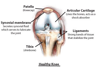
Healthy Knee

The end of your femur can be compared to a rocking chair. It has two distinct surfaces called compartments, which rest on the tibia. The third compartment is found behind the patella. All three compartments are covered with a tough lubricating tissue called cartilage.
Cartilage acts as a natural shock absorber, preventing bone on bone contact and providing a smooth, pain-free surface for the bones to glide against. The knee also contains synovial membranes, which produce synovial fluid to help lubricate and nourish the cartilage.
Links to Orthopedic Knee Education
Click on the links below for more educational information about the knee.
Links below are provided from the Orthopaedic connection website from the American Academy of Orthopaedic Surgeons.
Knee Arthritis
- Arthritis of the Knee
- Patellofemoral Arthritis
- Osteonecrosis of the Knee
- Osteotomy of the Knee
- Activities After a Knee Replacement
Treatment and Rehabilitation
All patient education materials are provided by OrthoPatientEd.com and have been reviewed by our Advisory Board of leading Orthopedic Surgeons to ensure accuracy. All materials are provided for informational purposes only and are not intended to be a substitute for medical advice from your orthopedic surgeon. Any medical decisions should be made after consulting a qualified physician.
This site includes links to other websites. OrthoPatientEd.com takes no responsibility for the content or information contained in the linked sites.
